Progress in building materials analysis (Part 1)


Summary: In this study two types of microscopes, a FEI XL-30 ESEM-FEG and a FEI Nova NanoSEM 230, were used which are capable of different high-resolution imaging techniques in order to characterize water containing and charging samples. In this field the environmental-SEM (ESEM) – technology in connection with high resolution electron microscopy is especially important, since water vapor can be used as gas in the sample chamber. Thus, water containing structures and phases can be investigated without sophisticated preparation
i. e. in their native states. Part 1 of the article summarizes the experimental setup and the optimization measures, such as low voltage SEM. The newly developed NanoSEM combined with Helix detector, cryo-SEM, EDS and EBSD capabilities offers various advantages.
The properties of cementitious binders such as workability, setting behavior, strength and durability depend directly on the hydration process. In order to understand the relations between material properties and material formation processes one has to understand the multiscale heterogeneous structure which is created. Water does not only play an important role in the hydration process but also with respect to the durability of cement-based and other building materials. Furthermore, water or pore solution are essential components within the microstructure of cementitious...
The properties of cementitious binders such as workability, setting behavior, strength and durability depend directly on the hydration process. In order to understand the relations between material properties and material formation processes one has to understand the multiscale heterogeneous structure which is created. Water does not only play an important role in the hydration process but also with respect to the durability of cement-based and other building materials. Furthermore, water or pore solution are essential components within the microstructure of cementitious systems representing the materialistic bracket over all observation levels – from the centimeter size grain down to the mesogel pore range of a few nanometers. Ultra high resolution FE-SEMs have the potential to visualize such structures continuously from macroscopic resolution down to the molecular level (seven magnitudes in the length scale). This wide range of magnification allows the study of nm-scale structures in the complex microstructure of building materials. However, the resolution does not depend on the primary beam diameter of the SEM alone, but it is also a function of complex beam-specimen interactions therefore the resolution depends on the nature of the specimen itself. In addition, specimen preparation is equally important in order to use the full potential of modern ultra high resolution SEMs.
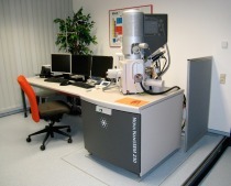
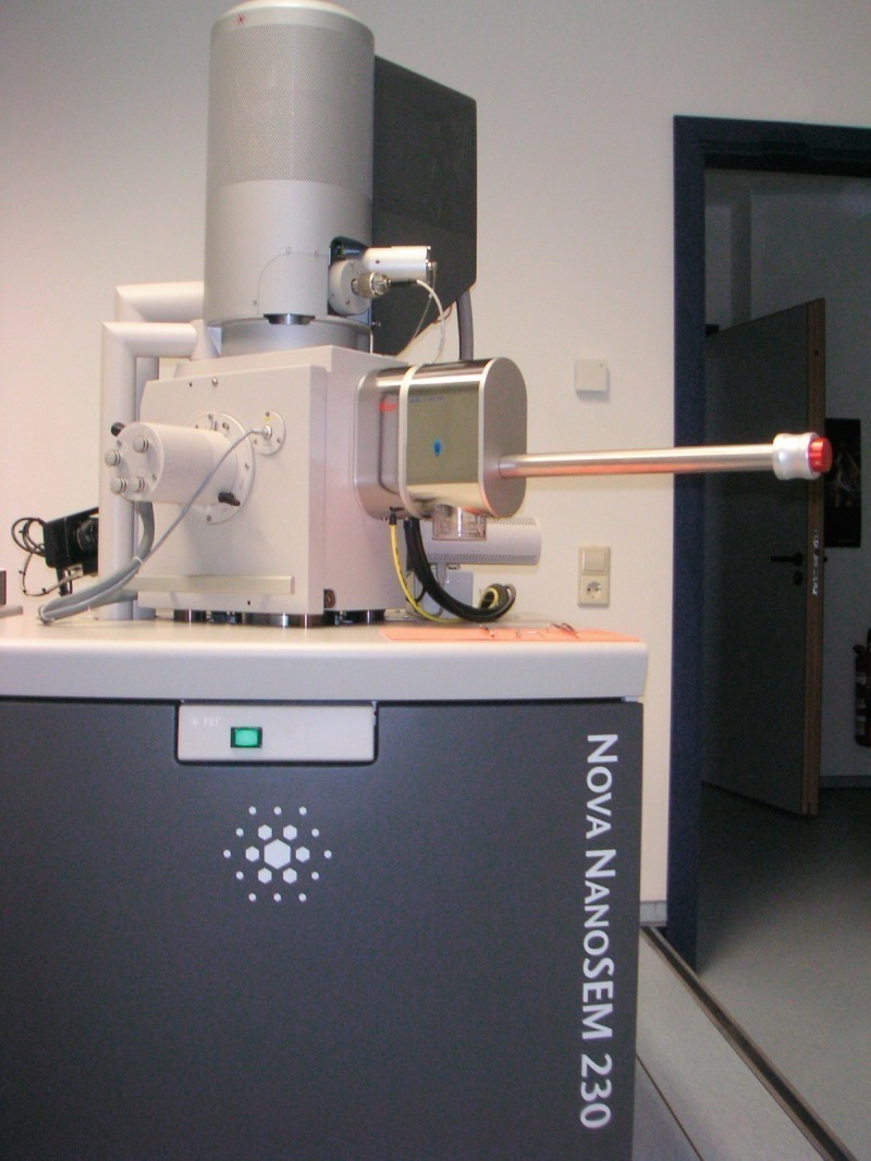
At the F. A. Finger Institute for Building Materials Science at the Bauhaus-University Weimar a new microscope has been recently set up (FEI Nova NanoSEM 230 (Fig. 1). Now two microscopes are available that are capable of different high-resolution imaging techniques in order to characterize water containing and charging samples. In this field the environmental-SEM (ESEM) technology in connection with high resolution electron microscopy is especially important, since water vapor can be used as gas in the sample chamber. Thus, water containing structures and phases can be investigated without sophisticated preparation i. e. in their native states.
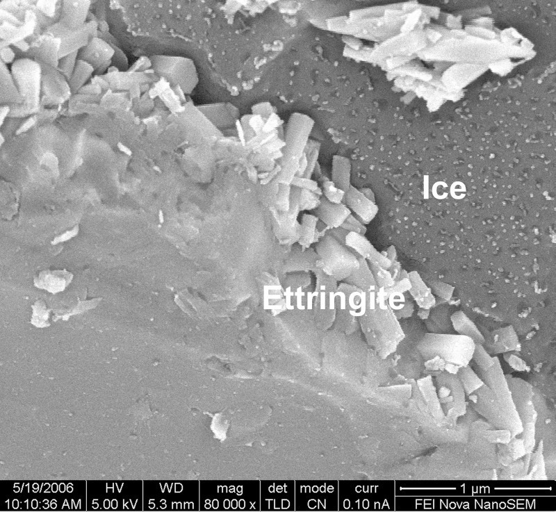
The ESEM-FEG can operate with a high water vapor pressure of up to 13.3 mbar. By means of a Peltier stage the observation of dynamic processes such as crystallization, dissolution, hydration etc. becomes possible. In addition in-situ investigations on samples which are exposed to micro-mechanical stresses can be carried out. In ESEM-WET mode (in which wet samples are investigated) there is a danger that amorphous and/or crystalline precipitation occurs and remains on the sample surface. These precipitations are created from the pore fluid in which different ions are solved. Therefore it is also important to produce a freshly fractured surface by means of a micromanipulator during the examination of the samples in the microscope.
The Nova NanoSEM 230 combines magnetic immersion lens technology with ESEM technology (Helix gaseous secondary electron detector). The combined effect yields unique ultra high resolution low vacuum characterization capabilities in a water vapor environment (gaseous mode) under low voltage conditions (smaller than 5 kV). Low energy electrons reduce beam penetration and optimize the amount of surface information carried by the signal. This microscope can also be used as a cryo-SEM for the investigation of high pressure frozen samples in high vacuum environment without coating or sputtering to avoid obscuring the nano-scale sample details of the hydrating structures. The cryo-SEM, the low vacuum (up to 1.6 mbar) as well as the ESEM methods (up to 13.3 mbar) have their own merits and are useful when they are used together. High resolution, low voltage SEM (LV-SEM – Helix detector) is also a powerful tool for studying very dense and compact microstructures (UHPC) as well as the nano-scale phases in the microstructure. Because LV-SEM provides images with a clear topographic contrast even on specimens containing phases with low density such as hydrate phases. Thus, the confusion of structural overlap does not occur. Furthermore, in microanalytical measurements the application of the LV-SEM mode reduces the excitation volume of the electrons up to a range of smaller than 500 nm and achieves a better excitation and X-ray emission for light elements.
The following sections illustrate, by means of some selected examples, how it becomes possible to increase the information depth by combining the different electron microscopic techniques and other physical methods. The method combination described here makes it possible to link morphological, chemical and structural material characteristics to each other. Clear statements can be made by combining the experimental possibilities and the physical characteristics of the individual methods. Preparation artifacts and beam damages which particularly occur in aqueous samples become obvious and measures to avoid them are described.
Triclinic C3S was synthesized by triple burning of a mixture of calcium carbonate (CaCO3, Merck, p. a.) and amorphous silica (SiO2, Merck, p. a.) at a temperature of 1,823 K (free lime content of C3S: 0.2 wt.-%). C3S was ground by means of a ball mill to a specific surface area of 4960 cm²/g (BET) and an average particle diameter of 9 µm (LS Coulter 230) was reached.
The cement was an OPC CEM I 42.5 R from Germany with a low (0.36 wt.-%) and a high (1.16 wt.-%) water soluble potassium oxide content (mainly potassium sulfate). Furthermore, the investigations were carried out on two UHPC reference mixtures (M2Q, B4Q; [1]). An OPC CEM I 52.5 R – HS/NA and the following additives and aggregates were used: silica fume, quartz powder, quartz sand (0.125/0.5 mm), basalt (2/8 mm), and superplasticizers (SP) based on polycarboxylate ether. In order to improve the ductility of the material steel fibers (length/diameter: 9.0/0.15 mm) were used. The chemical composition and the properties of the raw materials can be taken from [2].
Electron microscopy was carried out using two types of instruments an FEI XL30 ESEM-FEG and an FEI Nova NanoSEM 230 (FEI Company, Hilsboro, Ore., USA). The imaging by means of ESEM-FEG was carried out at high accelerating voltages between 15 and 30 kV. In order to investigate samples in the ESEM-WET mode at a relative humidity close to 100 % a Peltier stage must be used to cool the samples. A relatively high water vapor pressure in the sample chamber between 6.6 and 13.6 mbar was used.
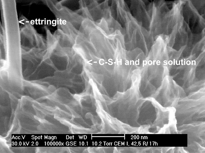
During the early hydration process of cementitious materials it is possible to image the microstructure in detail at high accelerating voltages (V0). With the hydrations process progressing it becomes clear, however, that the hydrate phases formed which have a comparably low density (1.7 g/cm3 for ettringite and approximately 2.2 g/cm3 for C-S-H phases) are strongly penetrated by the incident electrons at high accelerating voltages betweeen 20 and 30 kV which are necessary for high gas pressures in the sample chamber of the ESEM in the WET mode. Through crystal formation and growth during the hydration process the microstructure becomes more and more dense. In the ESEM-WET mode the high gas pressure in the sample chamber causes a significant scattering of the primary electron beam. The gas molecules scatter the electrons away from the high resolution focus. The scattering phenomenon has been labeled “beam skirting”. Furthermore, the amount of beam electrons that are scattered depends largely on the type of gas, the beam gas path length and the accelerating voltage. By increasing the beam energy the scattering effect becomes smaller (ceteris paribus) and the signal to noise ratio and thus the resolution improves. On the other hand the surface contrast gets almost completely lost and certain structure components cannot be imaged clearly although the resolution is high.
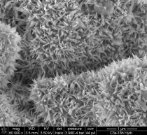
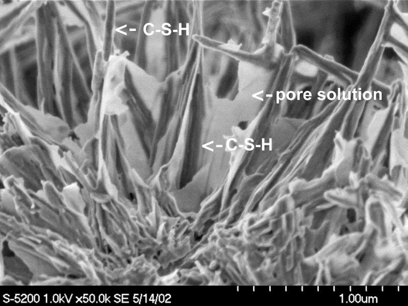
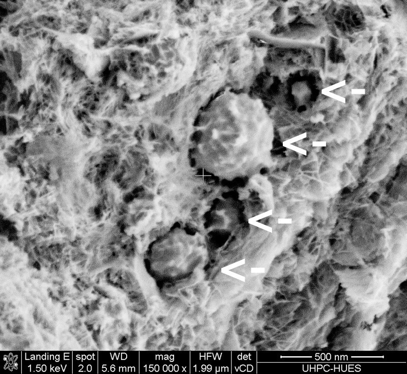
As shown in Figure 2a the structure components formed appear “diffuse, structurally overlapping” and not very high in contrast at high accelerating voltage conditions. In this case a detailed visualization of the microstructure is only possible using ultra high resolution imaging in the LV-SEM mode. At low V0 the penetration depth of the primary electron beam is reduced which means that the imaging-forming-signal is produced closer to the impact point. Pore solution covers the needle-like C-S-H phases and creates very thin films (thickness smaller than 10 nm) in the cement stone structure. Only by means of reducing of the primary beam voltage (smaller than 1.5 kV) it can be clearly distinguished between the C-S-H phases (diameter smaller than 50 nm) and the pore solution in the cement stone structure. The penetration depth of the electron beam at an accelerating voltage of 1 kV is only 23 nm (calculated for tobermorite). In doing so the C-S-H phases appear dark and the very thin pore solution films in the C-S-H structure appear bright (Fig. 2b). These thin films have such a tiny dimension that they are too small for elemental analysis by means of energy dispersive X-ray spectrometry (EDS). The proof that these thin films are pore solution indeed, can only be given indirectly by comparison of the microstructures of C3S hydrated with and without synthetic pore fluid (main component KOH). If one replaces such pore fluid by distilled water no such films occur [3].
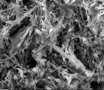
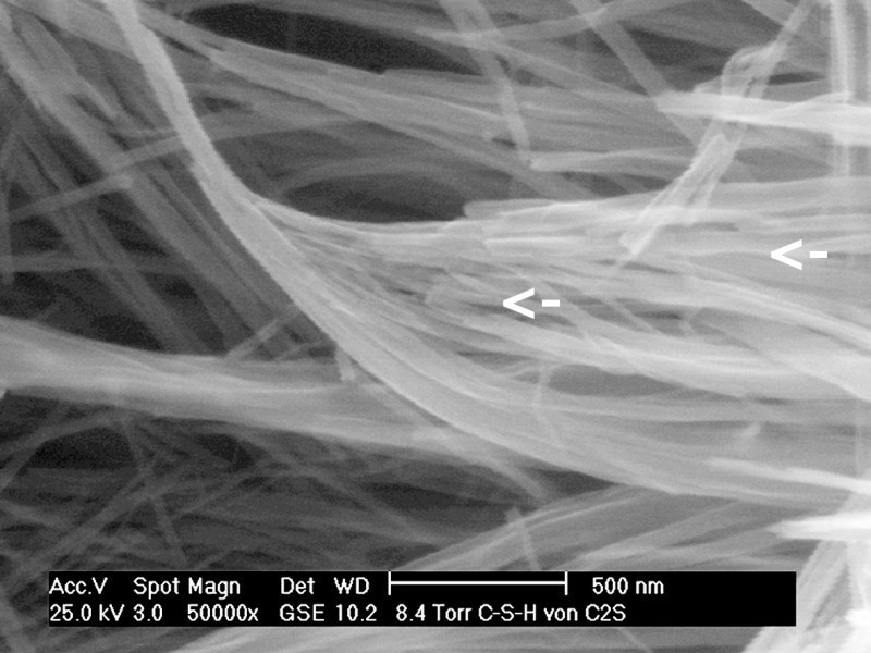
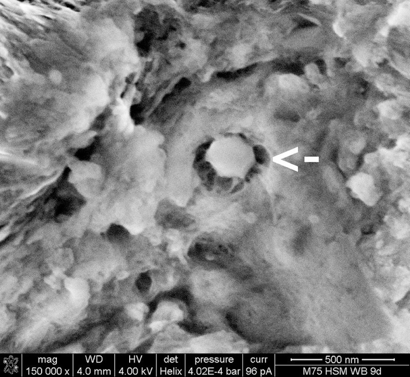
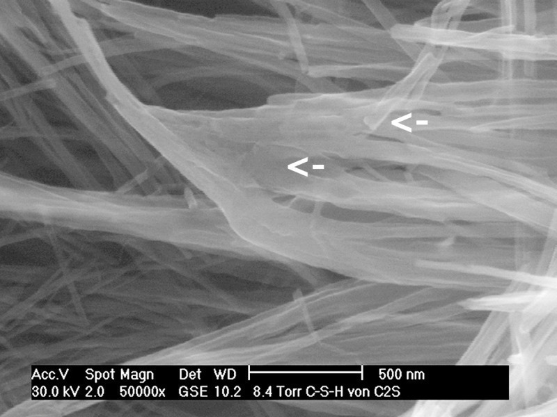
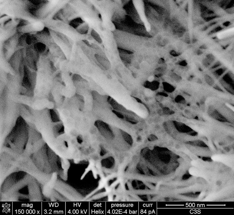
Like in the case of biological specimens in “Life Science” the achievable resolution depends not only on the microscope properties but it is also limited by beam damages (radiation damage at high charge density [4]). The comparison between the left and the right image in Figure 3 shows that it is possible that even the C-S-H phases are strongly damaged in ESEM-WET mode under high resolution conditions, especially when exposed to a high electron intensity (high probe current of 150 pA is equivalent to the charge density of 0.8 C/cm2 under these conditions). It is clearly visible that the parallel aligned C-S-H phases with a thickness in some cases of only 20 nm show a formation of an “indefinable mass” caused by beam damage. The rise of the specimen temperature results in an irreversible thermally induced damage.
This phenomenon is well-known from comparative investigations on the explanation of preparation artifacts which occur on samples prepared for the conventional SEM. Such artifacts can be the thermal radiation under vacuum conditions during evaporating carbon or heavy metal on the sample surface (coating or sputtering). This leads to such melting phenomena [5]. Particularly concerned are high and thin burrs or phases with fibrous habit (e. g. C-S-H phases).
Compared to the high-beam-energy investigations, high resolution LV-microscopy provides the following advantages: better secondary electron images (Fig. 2a and 2b as well as Fig. 4), higher electron spatial resolution, higher X-ray spatial resolution, reduced penetration of the electron beam, better X-ray emission for light elements, less damage as well as less charging of the sample. For these investigations a FEG-SEM (Nova NanoSEM230) with imaging capabilities in a water vapor pressure of up to 1.6 mbar in the sample chamber was deployed. High resolution (e. g. 1.8 nm at 3 kV on ideal samples such as gold on carbon) in low voltage mode without partial charge build-up can be achieved by means of a Helix detector when the magnetic immersion lens technology is combined with the ESEM technology. The resulting effect enables ultra high resolution, low-vacuum characterization capabilities. By means of this type of microscope it becomes possible to choose excitation conditions for non-coated and non-conductive samples in such a way that contrast rich imaging as well as spatial resolution in the microanalysis can be optimized. This method enables us to see structures under ultra high resolution conditions which are not visible using conventional SEM or ESEM.
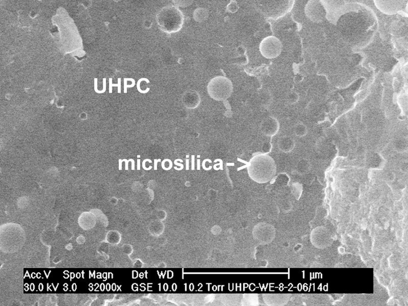
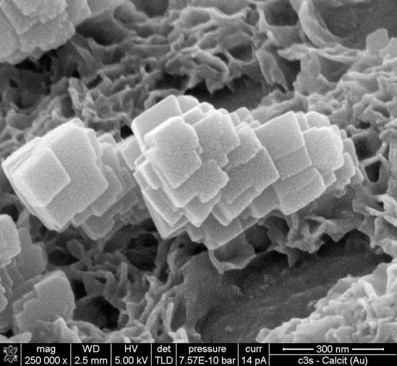
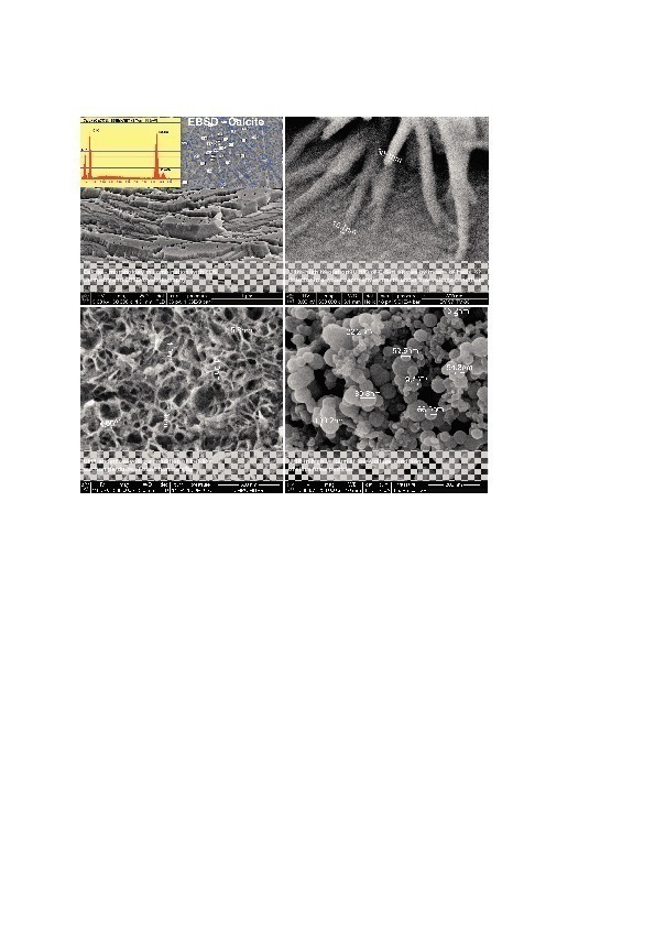
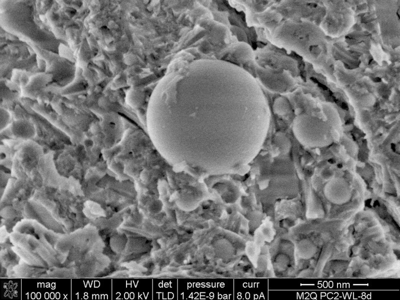
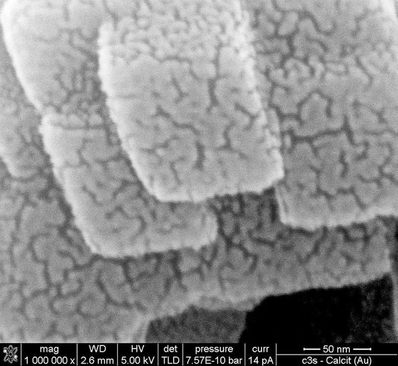
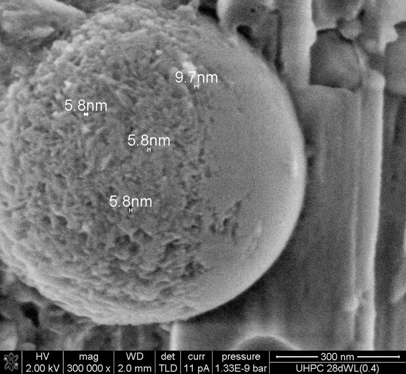
As the micrographs in Figure 4 show, microscopy with optimized electron energy is especially helpful when extremely dense structures for example ultra-high performance concrete (UHPC) is to be imaged. The best approach is to look at uncoated sample surfaces, because artifacts related to surface coating can be avoided (imaging by means of ESEM or Helix detector). Figure 5 shows that the NanoSEM can produce enlarged images of conductive or coated samples at low voltage conditions achieving magnifications of over 1000 000x and this contamination free and without vibration effects. The bad thing is, however, that we see the 2 nm gold coating layer structure but unfortunately not the original surface structure of the calcite crystals.
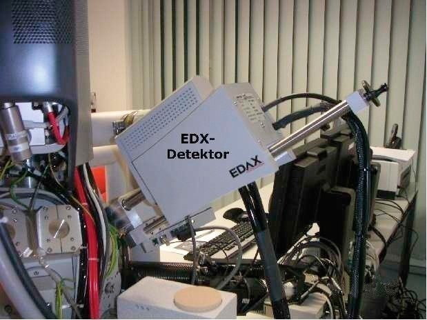
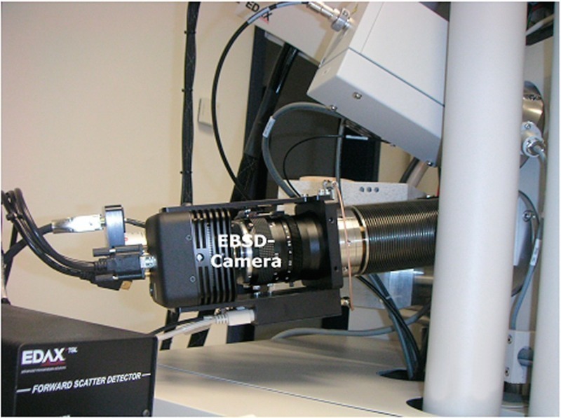
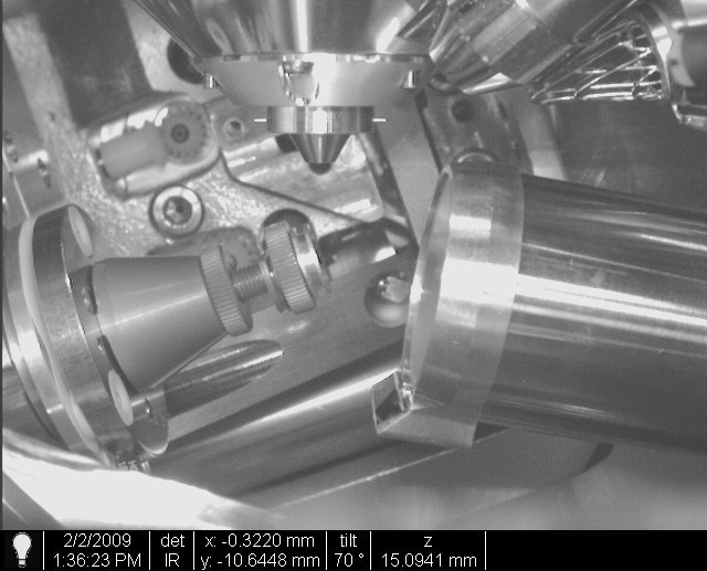
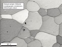
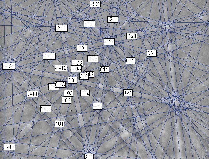
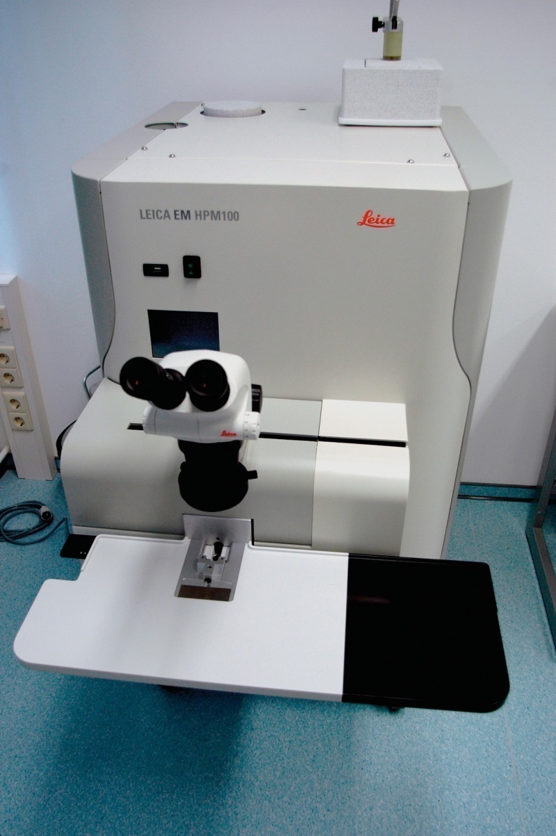
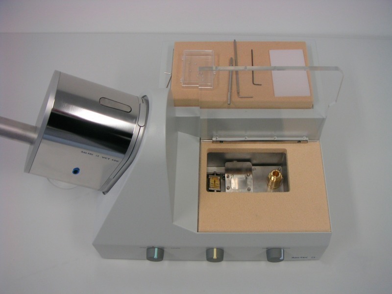
Furthermore, the Nova NanoSEM 230 is equipped with an analytical tool and a high-vacuum cryo transfer system. The analytical equipment (Pegasus XM4; EDAX Inc., USA) allows simultaneous data acquisition and analysis from any analytical point by means of energy dispersive spectroscopy (EDS: chemistry, Fig. 6) and electron backscatter diffraction (EBSD: crystallography; Figs. 7–10). This method combination allows a comprehensive material characterization. For the data acquisition by means of EDS an Apollo 40 silicon drift detector having an effective area of 35 mm2 is used. Thus, a high collection efficiency can be achieved. In addition, a high-vacuum cryo-transfer system (VTC 100) combined with a high pressure freezing machine (EM HPM 100) for the cryo-fixation (BAL-TEC now owned by Leica Microsystems, Figs. 11–13) was deployed.
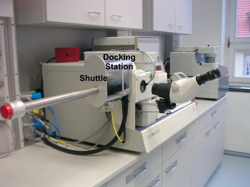
The cryo-fixation done by high pressure freezing (2100 bar) allows the fixation of native samples with a size of up to 200 µm thickness and 2 mm width. Minimal or even no ice crystal damage occurs, no chemical fixation or anti-freezing agents are necessary [6]. High pressure cryo-prepared samples (e. g. fresh cement pastes) can be used especially well for analytical EDS – EBSD investigations. The VTC 100 system allows autonomous sample cryo-preparation and SEM imaging. Both processes are linked through a high-vacuum cryo shuttle. Only an airlock (docking station, Fig. 14) is attached to the microscope in order to carry out the sample transfer into the SEM cooling stage. After the sample transfer has taken place the cryo shuttle can be removed from the docking station in order to achieve an absolute vibration free ultra high resolution imaging.
The application of improved and new imaging and analytical techniques enable a better and detailed characterization of the micro- and nanostructure of hardened cement pastes and give an improved insight in the microstructural development of cementitious material. In Part 2 of the paper answers to fundamental questions on cement chemistry will be presented.
Überschrift Bezahlschranke (EN)
tab ZKG KOMBI EN
This is a trial offer for programming testing only. It does not entitle you to a valid subscription and is intended purely for testing purposes. Please do not follow this process.
This is a trial offer for programming testing only. It does not entitle you to a valid subscription and is intended purely for testing purposes. Please do not follow this process.
tab ZKG KOMBI Study test
This is a trial offer for programming testing only. It does not entitle you to a valid subscription and is intended purely for testing purposes. Please do not follow this process.
This is a trial offer for programming testing only. It does not entitle you to a valid subscription and is intended purely for testing purposes. Please do not follow this process.